Answer: Coagulation! Isn't that exciting?!
But what is coagulation you may ask? Well, to be more correct we could say blood coagulation, which is essentially the formation of a blood clot. You know when you get a cut and then your body heals itself and sometimes there's a scab? Some of you might even have a faint recollection of the word "platelets" from your early biology days. But I suppose I'm getting rather ahead of myself.
And actually, now that I really think about it--coagulation is only a part of what we will be learning; a nonetheless useful part that, when combined with others that will be discussed, will paint the picture of the Hemostasis System. And I realize I may have lost some people, but stick with me for another moment and I'll fix it. (for some reason this whole unit is called Coagulation even though it should technically be Hemostasis and I've just now come to this realization. Professors can make things so much more difficult than they need to be at times, don't they?)
To make things a little easier, here is a basic outline of what I will be discussing:
HEMOSTASIS
- Primary Hemostasis
~ Platelets
- Secondary Hemostasis + Fibrinolysis
~ Coagulation Cascade
- Disorders
~ Platelet Disorders
~ Coagulation Disorders
~ Fibrinolytic Disorders
~ Thrombosis/Thrombophilia
- Anticoagulant Therapy
That doesn't seem too bad now, does it? It can even be boiled down to:
"The Hemostasis System and its Disorders"
or even:
"How Hemostasis Works and What Can Go Wrong"
or even simpler and in more colloquial terms:
"The Way Your Body is Supposed to Heal Itself After a Cut and Different Ways it Can Mess Up & Screw You Over"
--because where's the fun in having your body do exactly what it's supposed to? Every body has their dysfunctional part and just be grateful bleeding isn't yours.
MOVING ON! I promise all of this isn't going to be in one post--I'm not that mean nor do I wish to inflict that much pain upon myself. There will be a number of posts, set up as sections similar to the ones in my first outline.
Now before we start talking about Primary Hemostasis, how about I take some time to teach you the basics of just what hemostasis is? Was this what we learned the first day of this unit? Yes. Now it'll be just like you were there with me three Thursdays ago at 8 in the morning; though please consider yourselves lucky because you have me here to teach it to you in a manner in which you can actually understand it the first time through.
 So--hemostasis is defined as a localized and controlled process that maintains blood fluidity and fixes it upon injury. It's made up of three components.
1. Platelets
2. Vascular components
3. Coagulation Proteins
These three parts interact in various ways to bring about the different steps of hemostasis:
1. Primary hemostasis
- The vascular and platelet response to vessel injury
2. Secondary hemostasis
- Response of the coagulation process to vessel injury
3. Fibrinolysis
- Gradual clot dissolution (uses all three)
That's about how in depth I'll get about that--they'll all be discussed further in the later posts. For further preparation though, how about a review of blood vessel structure (vascular) and some vocabulary?
First off, all blood vessels share a similar structure with a central cavity (lumen) that the blood flows through. The lumen is lined with a single layer of endothelial cells that protect both the cellular elements of the blood that flows through the lumen and then the surrounding tissues. There are three blood vessels: arteries, veins, and capillaries.
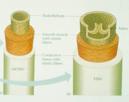
1. Tunica adventitia
- made up of external elastic tissue, loose connective tissue with collagen fibers, and fibroblasts
2. Tunica media
- made up of smooth muscle cells and elastic tissue
- this allows it to contract and expand, so it's responsible for vasoconstriction and vasodilation, which occur in primary hemostasis
3. Tunica intima
- made up of a single endothelial cell layer, the basement membrane and internal elastic lamina composed of elastic connective tissue
Capillaries then, act as a connection between the veins and arteries, and is the site where metabolic exchange between the blood and tissues take place. Their structure is smaller and less complex than the other two, but no less important. They're composed of a single endothelial layer and compose the largest surface area.
Time for some vocabulary:
Basic Definitions
- Hypocoagulation--bleeding
- Hypercoagulation--thrombosis, inappropriate formation of thrombi in the vasculature that occlude normal blood flow
- Coagulopathy--disorder of blood coagulation
- Bleeding diathesis--predisposition to bleeding
Bleeding manifestations

- Petechiae--pinpoint hemorrhages
- Purpura--larger than petechia but smaller than ecchymosis, a collection of petechiae
- Ecchymosis--spread of blood within a single layer, bruise
- Hematoma--localized collection of blood outside the vessels
- Hermarthrosis--bleeding in the joint
- Hematuria--blood in the urine
Hypercoagulation manifestations
- Thrombus--clot formation in moving stream, intravascular rather than extravascular
- Embolism--clot that has moved from original site of formation
And that's it for now! Does it make sense? You with me so far? Good. Onward to primary hemostasis!
No comments:
Post a Comment